Surface Anatomy
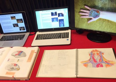
This work was presented by Frederick, Nydile and Amy at a UoB/Bristol Museum and Art Gallery event ‘Leonardo: Between Art & Medicine’ led by the UoB History of Art Department in April 2019. The event included presentations from diverse disciplines, and ‘Surface Anatomy’ was later shared in a temporary exhibition alongside the touring ‘Leonardo’s Drawings’. Following informal viewing and discussion the students reported that members of the public asked predominately about having a mobile app which would employ a similar concept as ‘Surface Anatomy’ allowing them to ‘see’ into their bodies and observe particular structures in detail. They also thought this could be relevant educationally across generations and communities.
Group 19
These creative pieces were part of the annual collaborative presentations at the Year One Foundation of Medicine Conference, 2018.
Other group members included: Nicolas James Pugh, Ellie Pybus, Mariam Rama Javed, Alexandra Razumovskaya-Hough, Millicent Richardson, Renan Rivero, Niamh Roberts and Michaela Margaret Rogers.
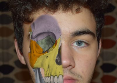
This piece really stood out me, I feel that often the complexities of the body can be overlooked from an outsiders view; whilst conversing or looking at an individual. This piece shows how there may need to be more appreciation for the processes and anatomy of the human body, with the use of art enabling this.
This piece demonstrates the intricacy and wonders of the human body. We complete day to day tasks and often do not realise or even think about how we are able to do these things. As I type this review now it is crazy to think of the amount of work the body is currently doing to allow me to do this. This piece helps capture the delicate and vast configuration of the human body.
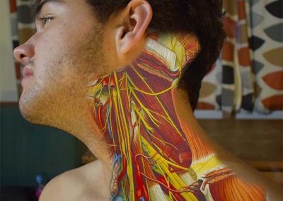
‘Surface Medicine’ is a piece of medical art that superimposes medical anatomical images onto individual anatomy. This creates an almost surrealist style which is both profound and aesthetically pleasing to observe.
The artwork was submitted to the ‘Leonardo: Between Art & Medicine’ event at Bristol Museum and Art Gallery in 2019. In my opinion, it serves as a perfect continuation of Leonardo da Vinci’s style of anatomical sketches into the modern era. It is both evidently different yet also appears stylistically similar to the pieces that inspired it.
The vibrantly coloured, twisted patterns of the anatomical features such as blood vessels, nerves, and bones create a simultaneously educational, beautiful, yet also somewhat unsettling feeling within the observer. The fact this piece can evoke such a broad spectrum of emotions is another reason I selected it for my review.
It also evokes a sense of awe and personal connection towards my own anatomy when I view it, as seeing the complexities of the human body, superimposed upon images of people, serves as a reminder that although I cannot see them, these anatomical features are also within me.
Due to this artwork combining the beauty of art with the complexity of medicine, as well as perfectly continuing its stylistic inspiration’s vision and style, this is my favourite piece of art submitted to the ‘Out of our Heads’ exhibition.
This is interesting because it links what we have just learned and it an interesting way of visualising what happens beneath the skin which we cannot see.
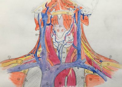
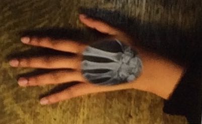
This piece really stands out to me for 2 reasons, namely, the immense detail and the inspiration behind the piece. To start off, the set of 3 images, integrating the segments of scan with external surface of the human body, are in great detail. They show the individual bone segments, nerves, and blood vessels, of the various body areas they overlie- these include the subclavian vein in the neck, the metacarpals in the hand, or orbitals in the skull. Such detail allows the viewer to appreciate the intricate mechanisms that make up the human body, and by extension, the beauty of it. Moreover, the motivation behind the piece (the group member with scoliosis) was another salient aspect of the piece. This was connected to the mention regarding the development of an app, that can produce similar images. These images would allow patients to gain knowledge about the anatomy of their condition- it connects to the GMC guidance of giving patients information they need/want in a way they understand. Hence, they might make more informed decisions about their treatment, in the future. To conclude, the detail of the images, and the inspiration behind the image, brought out this image.
This piece caught my attention as it emphasizes the sheer complexity of the human body masked by the relatively unassuming appearance of the skin. It not only highlights the intricate details of the underlying structures within us but also evokes the sense that we should appreciate the differences between each of us that are not apparent to the naked eye—this is not limited to anatomical variants but also characteristics like neurodivergence and invisible disabilities.
The motive for creating a mobile app with “see-through” functions is an excellent idea to empower patients and, for those interested in learning more about themselves, to gain more knowledge- especially if relevant to their conditions or simply to satisfy one’s curiosity.
I like this piece because it gives the layman a visual representation of what happens beneath their skin, and it shows that there is a lot of more to people than meets the eye (what is on the surface). It emphasises the complexities of the human body and gives an insight into just how much is happening in the body at the same time.